Ultrasound-guided fine needle aspiration and surgeon-performed ultrasound
Ultrasound guided FNA
Ultrasound guided fine needle aspiration is a minimally invasive procedure performed on the head and neck area to investigate thyroid nodules, lymph nodes, salivary gland masses or other head and neck lumps. In this procedure, cells are removed from the area of investigation and sent for testing.
Our surgeons can perform ultrasound-guided fine-needle aspiration (US-FNA) on the same day as a consultation, saving multiple appointments, costs, travel and stress for patients.
What is involved in ultrasound guided fine needle aspiration?
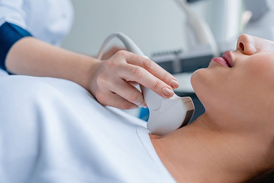
A fine needle aspiration is a short procedure taking approximately 10 minutes. During the procedure an ultrasound machine is used to locate the area to be sampled while a very thin needle is inserted. The needle is then moved gently in the area under ultrasound imaging to take a sample of cells before being removed. The procedure is followed by a short recovery period before going home.
There is no fasting or other preparation required. A local anesthetic may be used to numb the area prior to the procedure.
Surgeon Performed Ultrasound
Ultrasound is a highly operator-dependent investigation. A surgeon-performed ultrasound involves the specialist surgeon performing a dedicated ultrasound on the patient in the office or consultation room.
This allows the treating specialist to perform a surgical-oriented scan, looking for features that are important from the surgical point of view (and sometimes not addressed on conventional scans).
A surgeon-performed ultrasound permits the surgeon to clearly display scan findings to the patient in real-time, providing a description within the clinical context, so that the patient is better informed. It avoids radiation and is safe and accurate.
Conditions best suited for surgeon-performed ultrasound
Surgeon-performed ultrasound is extremely useful for the workup of patients with benign neck lumps, parotid and submandibular gland masses, head and neck cancer, thyroid nodules and parathyroid adenomas.
When combined with US-FNA, it is the most accurate imaging tool in the detection of pathological lymph node metastases in the head and neck. It is performed by otolaryngology head and neck surgeons throughout North America, Asia, Europe and Australia and has been shown to change the surgical management in a large percentage of patients.


