Ear Conditions
Otitis Externa (Swimmer’s Ear)
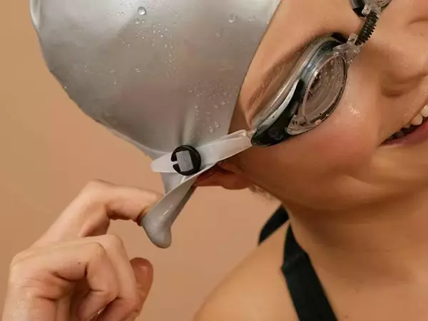
Swimmer’s ear or otitis externa is an infection or inflammation of the outer ear canal — the passage between the outer ear and the eardrum. Commonly known as swimmer’s ear, it is a painful but highly treatable condition that affects people of all ages. ENT Specialists Group offers swimmer’s ear treatment for children and adults.

Medically reviewed by Dr Yi Chen Zhao, FRACS, ENT Surgeon
Last reviewed 20 August 2026.
What is Otitis Externa?
Otitis externa is an infection or inflammation of the skin lining the ear canal — the tube that runs from the outer ear to the eardrum. Unlike middle ear infections (otitis media), otitis externa does not involve the middle ear or eardrum itself, but rather the external ear canal skin and surrounding soft tissue.
The condition is commonly referred to as swimmer’s ear because water trapped in the ear canal after swimming is one of the most frequent triggers. However, otitis externa can develop in anyone — swimmers and non-swimmers alike — as a result of minor trauma, skin conditions, or changes in the ear canal’s natural protective environment.
Most cases of otitis externa are acute and resolve within a week or two with appropriate topical treatment. A smaller subset of patients experience chronic otitis externa — a persistent or recurrent inflammation that can be more challenging to manage and often has an underlying dermatological or anatomical cause. In rare cases, particularly in older adults or those with diabetes, a severe form called malignant (necrotising) otitis externa can develop and requires urgent specialist care.
Contact Us
467 Upper Heidelberg Road,
Heidelberg Heights, VIC 3081
03 9998 7459
Recognising Otitis Externa
Symptoms typically develop over one to two days and can range from mild irritation to severe pain. The ear canal may become so swollen that hearing is temporarily reduced.
Ear Pain
Often the most prominent symptom — ranging from mild discomfort to intense, throbbing pain that worsens when the outer ear is touched or pulled.
Tragal Tenderness
Pain on pressing the small cartilage flap (tragus) at the entrance of the ear canal — a hallmark sign that distinguishes otitis externa from middle ear infection.
Itching
Intense itching inside the ear canal is common, particularly in the early stages and in cases with an underlying skin condition such as eczema.
Discharge
The ear canal may produce a clear, watery, or purulent (pus-like) discharge. An unpleasant odour may accompany bacterial infection.
Reduced Hearing
Swelling of the ear canal walls or a build-up of discharge and debris can partially block the canal, causing temporary muffled hearing.
Redness & Swelling
The skin of the ear canal appears red and swollen. In severe cases, swelling may extend to the outer ear (pinna) and surrounding skin.
When to seek urgent care
If you are experiencing spreading redness or swelling beyond the ear, fever, severe pain not responding to analgesia, facial weakness, or symptoms in a person with diabetes or a compromised immune system, seek urgent care. These may indicate necrotising (malignant) otitis externa — a serious condition requiring urgent specialist assessment.
What Causes Otitis Externa?
The ear canal is naturally protected by a slightly acidic environment and a thin layer of cerumen (earwax) that traps debris and resists bacterial and fungal growth. Otitis externa develops when this protective barrier is disrupted, allowing infection or inflammation to take hold.
Water Exposure
Prolonged or repeated exposure to water — particularly in swimming pools, lakes, or the ocean — softens and macerated the ear canal skin, disrupts its acid mantle, and creates a warm, moist environment ideal for bacterial growth.
Bacterial Infection
The majority of acute otitis externa cases are caused by bacteria, most commonly Pseudomonas aeruginosa and Staphylococcus aureus. These organisms thrive once the skin’s natural defences are compromised.
Skin Conditions
Underlying dermatological conditions — including eczema, psoriasis, seborrhoeic dermatitis, and contact dermatitis — can affect the ear canal skin, leading to chronic otitis externa that is driven by inflammation rather than infection alone.
Fungal Infection (Otomycosis)
Fungal otitis externa — usually caused by Aspergillus or Candida species — is less common but important to identify, as it requires antifungal treatment rather than antibiotics. It is more frequent following prolonged antibiotic ear drop use.
Narrow or Hairy Ear Canals
Anatomical variations that reduce natural ventilation or impede earwax migration — such as a narrow or tortuous canal — can predispose to moisture retention and recurrent infection.
Trauma to the Ear Canal
Scratching the ear with fingernails, cotton buds, hearing aid components, or earphones damages the delicate canal skin, creating entry points for infection and stripping protective earwax.
Hearing Aids & Earphones
Regular use of in-ear devices can trap moisture, reduce ventilation, cause minor trauma, and introduce bacteria into the canal. People who wear hearing aids have a higher rate of otitis externa.
Diabetes & Immunosuppression
Poorly controlled diabetes and immunocompromised states significantly increase the risk of severe otitis externa, including the rare but dangerous necrotising (malignant) form caused by Pseudomonas spreading into surrounding bone and soft tissue.
Not Just a Swimmer’s Problem
Despite the nickname, otitis externa is far from exclusively a condition of swimmers. Anyone who uses cotton buds, wears in-ear hearing aids or earphones, has eczema-prone skin, or simply lives in a humid climate can develop the condition. In Australia’s warm summers, otitis externa is one of the most common presentations to general practice and ENT clinics — and recurrent or chronic cases are frequently under-treated without specialist input to identify the underlying cause.
How Is Otitis Externa Diagnosed?
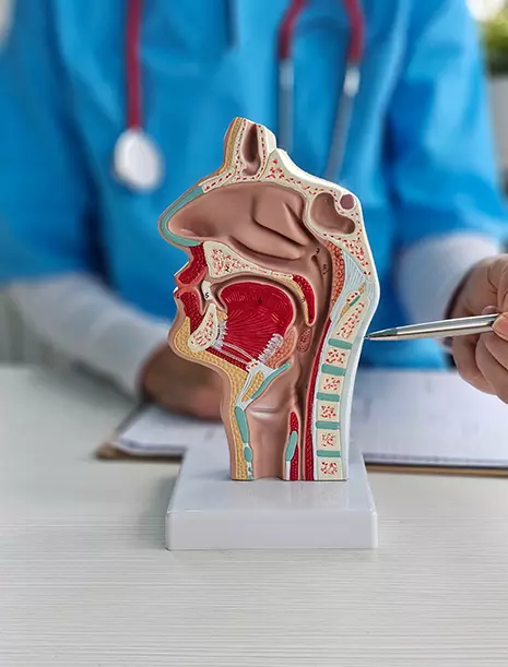
Diagnosis is primarily clinical. Your ENT specialist will examine the ear canal directly and, where appropriate, take swabs to identify the causative organism and guide targeted treatment.
Clinical History
Your specialist will ask about the onset and character of symptoms, recent water exposure, ear hygiene habits, use of in-ear devices, skin conditions, and any previous episodes. A history of diabetes or immunosuppression is also important to establish.
Ear Swab — Selected Cases
A swab of the ear canal discharge is sent for bacterial and fungal culture, particularly in cases that have not responded to initial treatment, are recurrent, or where fungal infection (otomycosis) is suspected. Identifying the specific organism and its antibiotic sensitivities allows targeted therapy.
Otoscopy and Microsuction
The ear canal is examined using an otoscope or operating microscope. The canal skin is assessed for redness, swelling, and discharge. Microsuction — gentle suction under magnification — may be used to clear debris and discharge from the canal, both to aid diagnosis and to allow ear drops to reach the affected skin more effectively.
CT Scanning (Selected Cases)
In cases where necrotising otitis externa is suspected — particularly in diabetic or immunocompromised patients with severe or persistent symptoms — a CT scan of the temporal bone is performed to assess for bony involvement and the extent of spread to surrounding structures.
Treatment Options
The majority of otitis externa cases respond well to topical treatment and resolve within one to two weeks. Recurrent or chronic cases, fungal infections, and severe presentations require a more tailored approach. Keeping the ear dry throughout treatment is essential regardless of the management pathway.
Topical Antibiotic Ear Drops
The mainstay of treatment for bacterial otitis externa. Drops containing an antibiotic — often combined with a corticosteroid to reduce inflammation — are instilled into the ear canal several times daily for one to two weeks.
Ciprofloxacin-based drops are commonly used due to Pseudomonas coverage
Combination antibiotic-steroid drops reduce pain and swelling more quickly
The ear canal must be clear of debris for drops to be effective
Drops should be warmed in the hand before instillation to reduce dizziness
Microsuction & Aural Toilet
Careful cleaning of the ear canal under microscopic visualisation removes discharge, debris, and shed skin that would otherwise prevent ear drops from reaching the infected canal wall. This is performed in clinic by your ENT specialist and often provides immediate pain relief.
Essential for moderate to severe cases
Allows direct inspection of the eardrum and canal
May need to be repeated at follow-up if the canal re-fills
Ear Canal Wick
When the ear canal is significantly swollen and ear drops cannot penetrate adequately, a small medicated wick (Pope wick or ribbon gauze) is gently inserted by a specialist to hold the canal open and deliver medication directly to the inflamed skin.
Used when swelling prevents drop penetration
Usually removed or replaced after 24–48 hours
Provides rapid reduction in canal swelling
Antifungal Treatment
Fungal otitis externa (otomycosis) requires antifungal ear drops or solution rather than antibiotics. Thorough aural toilet to remove fungal debris is a critical part of treatment, as antifungals cannot penetrate the fungal mat that forms in the canal.
Clotrimazole solution is the most commonly used topical antifungal
Treatment duration is typically two to four weeks
Recurrence is common if the canal is not kept scrupulously dry
Oral Antibiotics
Systemic antibiotics are not required for most cases of otitis externa, which respond well to topical therapy. They are reserved for cases with spreading cellulitis beyond the ear canal, significant systemic illness, or where topical treatment is not possible.
Used when infection spreads to the outer ear or surrounding skin
Antibiotic choice guided by swab culture results where available
Always required in necrotising otitis externa, often intravenously
Management of Necrotising Otitis Externa
This rare but serious form requires urgent hospital admission, prolonged intravenous antipseudomonal antibiotics, and close monitoring. Surgical debridement may be necessary if infection spreads to bone or surrounding structures.
Predominantly affects diabetic or immunocompromised patients
CT and nuclear medicine imaging used to monitor treatment response
Treatment duration often extends to six weeks or more
Multidisciplinary management with ENT, infectious diseases, and endocrinology
Keeping Otitis Externa at Bay
For people prone to recurrent episodes, a few straightforward habits can significantly reduce the risk of future infections.
Keep ears dry after swimming
Tilt the head to each side after swimming to allow water to drain. A hair dryer on the lowest, coolest setting held at arm’s length can help dry the canal gently.
Use ear plugs when swimming
Custom-moulded or well-fitted silicone ear plugs are effective at preventing water entering the ear canal — particularly useful for regular swimmers or those with a history of recurrent episodes.
Avoid cotton buds and ear probing
The ear canal is self-cleaning. Cotton buds remove protective earwax, traumatise the canal skin, and push debris towards the eardrum — all of which increase the risk of infection.
Acidifying ear drops (prophylactic)
For frequent swimmers, dilute acetic acid (vinegar) drops or commercial acidifying drops instilled after water exposure can help restore the ear canal’s natural acid environment and reduce bacterial colonisation.
Manage underlying skin conditions
If eczema, psoriasis, or seborrhoeic dermatitis is contributing to chronic otitis externa, optimising management of the underlying skin condition — with the help of your ENT or dermatologist — can significantly reduce recurrence.
Keep hearing aids clean and dry
Regular cleaning of hearing aid moulds and using a drying kit overnight can reduce moisture and bacterial build-up — an important step for hearing aid wearers prone to ear canal infections.
Frequently Asked Questions
No — swimming should be avoided until the infection has completely resolved, which typically takes one to two weeks. Continuing to expose the ear to water will prevent healing and is very likely to prolong or worsen the infection. Once you have recovered, using well-fitted ear plugs and drying the ears thoroughly after every swim can help prevent recurrence.
The key distinguishing feature is the location of the infection. Otitis externa affects the outer ear canal — the skin between the outer ear and eardrum — and typically causes pain that is worsened by touching or pulling the outer ear. A middle ear infection (otitis media) occurs behind the eardrum and usually follows a cold or upper respiratory infection, causing deep-seated ear pain and sometimes fever. Both conditions can cause hearing reduction, but they are managed very differently.
The most common reason ear drops fail to work is that the ear canal is blocked by debris, discharge, or swelling — preventing the drops from reaching the infected skin. An ENT specialist can perform microsuction to clear the canal so that drops can penetrate effectively. In some cases, the causative organism may be resistant to the antibiotic in the prescribed drops, or the infection may be fungal rather than bacterial, requiring a different medication entirely.
Chronic otitis externa is persistent or recurrent inflammation of the ear canal lasting more than three months. It is often driven by an underlying cause such as eczema, contact allergy to ear drop preservatives or hearing aid materials, seborrhoeic dermatitis, or habitual ear canal trauma. Identifying and addressing the underlying trigger — rather than repeatedly treating acute flares with antibiotics — is the key to long-term resolution.
Despite its alarming name, “malignant” here refers to the aggressive behaviour of the infection — not cancer. Necrotising (malignant) otitis externa is a rare but serious extension of bacterial otitis externa into the bone of the skull base, almost exclusively affecting people with diabetes or severely compromised immune systems. It is characterised by severe, disproportionate pain, cranial nerve involvement (particularly facial weakness), and failure to respond to standard treatment. It requires urgent specialist assessment, hospital admission, and prolonged antibiotic therapy.
Mild, straightforward cases are often managed successfully by a GP with a short course of antibiotic ear drops. Referral to an ENT specialist is recommended when symptoms are severe, the ear canal is significantly swollen, there has been no improvement after an initial course of treatment, the infection is recurrent, fungal infection is suspected, or the patient has diabetes or is immunocompromised. Specialist review allows thorough aural toilet, targeted microbiological assessment, and identification of any underlying contributing factors.
Suffering from a painful or recurrent ear infection?
Our ENT specialists can assess, treat, and help prevent recurrent otitis externa — including thorough aural toilet, targeted treatment, and management of underlying causes.
Our ear nose and throat specialists will take the time to explain the process to you to ensure the best outcome for your little one.
Our surgeons are also members of the Australian Society of Otolaryngology Head and Neck Surgery.
If you’d like to understand more about treatment and surgical options for your child, contact us today or make an online booking.